
TSRA Content:
Author: Jessica G.Y. Luc, MD
This is a revision and update from the previous edition of the TSRA Primer in Cardiothoracic Surgery written by Zubair Hashimi, MD, and Jeremiah Martin, MD.
When considering coronary artery anatomy, it is helpful to visualize the system as a "ring and loop" (Figure 1). The ring is situated in the plane of the atrioventricular groove and consists of the right coronary artery (RCA) and the left circumflex coronary artery (sometimes abbreviated as the circ or Cx). The loop arises almost perpendicularly from this in the interventricular plane and consists of the left anterior descending (LAD) and the posterior descending artery (PDA). There are multiple possible points for bypass graft anastomosis between these territories. Keeping this three-dimensional framework in mind will assist you in interpreting coronary angiography.
The coronary artery system originates from the aortic root with the left and right coronary arteries. The left main coronary artery originates from the left coronary sinus of the aorta and courses between the pulmonary trunk and the left atrial appendage. The left coronary artery then divides into the LAD artery and the circumflex artery, with an occasional ramus intermedius artery in the middle of these two branches. The LAD courses in the anterior atrioventricular (AV) groove towards the apex of the heart. The LAD has septal perforator branches towards the AV groove and diagonal branches away from the AV groove. The LAD has been described in segments as proximal, mid, and distal. The proximal LAD is from its origin to the first diagonal branch or septal perforator. The mid LAD is from the origin of the first diagonal branch or septal artery to the origin of the last diagonal branch. The distal LAD is from the last diagonal branch to the end of the LAD. On angiography, the LAD can be reported as types 1-4. A type 1 LAD terminates before the apex. A type 2 LAD dually supplies the apex along with the right coronary artery (RCA). A type 3 LAD supplies the entire apex. A type 4 LAD wraps around the apex. The LAD supplies the anterior part of the septum, anterior wall of the left ventricle, and the atrioventricular bundle.
The circumflex artery travels in the left AV groove between the left atrium and left ventricle. The circumflex artery has branches including the obtuse marginal (OM) arteries which supply the lateral left ventricular wall. The circumflex artery terminates before the posterior interventricular groove in 80-85% and in 15-20% will continue as the PDA.
The RCA originates from the right coronary sinus of the aorta and courses along the right AV groove between the right atrium and the right ventricle to the inferior part of the septum.
The first branch of the RCA is a small conus branch that supplies the right ventricular outflow tract. The second branch is the sinoatrial (SA) nodal artery (in the majority of patients) which supplies the SA node. Next, several branches supply the anterior wall of the right ventricle. A large acute marginal branch comes off with an acute angle and runs along the right ventricle and diaphragm. RCA then continues and branches, giving off the PDA (80-85% of patients) that supplies the inferior wall of the left ventricle and the inferior part of the septum. An additional branch of the RCA that is more lateral and closer to the circumflex is the posterolateral (PL) branch. "Dominance" is determined by the artery (either right coronary artery or left circumflex artery) that supplies the PDA.
Interpreting coronary angiograms
Projections defined as right anterior oblique (RAO) and left anterior oblique (LAO) refer to the position of the image intensifier with respect to the patient. A good rule of thumb when trying to figure out the projection used in a view is to identify the spine. If the spine is on the right side of the screen, it is a LAO projection; if the spine is on the left, then it is a RAO projection; if the spine is midline then it is an anteroposterior (AP) projection. In addition, the projection can be tilted either cranially or caudally to better visualize certain vessels. For example, the LAO caudal view (spider view) is often used to better visualize the distal left main and proximal vessel anatomy of the left coronary artery.
Remember:
1. Two orthogonal views are always required to characterize the degree of stenosis of a lesion; the assessment is derived from the projection with the greatest narrowing
2. Always look at the complete catheterization series before coming to conclusions about stenosis.
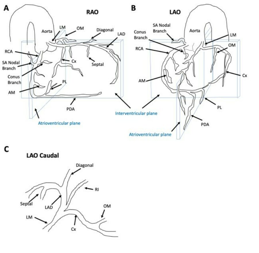
Figure 1: Views of the coronary artery system in the (A) right anterior oblique (RAO), (B) left anterior oblique (LAO), and (C) LAO caudal view. Abbreviations: AM, acute marginal artery; Cx, circumflex artery; LM, left main artery; OM, obtuse marginal artery; PDA, posterior descending artery; PL, posterolateral artery; RCA, right coronary artery; RI, ramus intermediate artery; SA, sinoatrial artery.