
TSRA Content:
Author: Georgia R. Layton, MD
Intra-aortic balloon pumps (IABPs) are a form of mechanical circulatory support commonly utilized both pre- and post-operatively for cardiac surgery patients. They work to reduce myocardial oxygen demand and increase myocardial blood supply. IABPs are relatively cheap and easy to insert and can be managed in high-dependency or intensive care ward environments. Their benefit is modest compared to alternative circulatory support such as a ventricular assist devices, but they offer a good short-term solution for patients with compromised coronary circulation and/or cardiogenic shock.
An IABP sits in the descending thoracic aorta and works to decrease ventricular afterload, positively augment diastolic pressure and subsequently improve end-organ perfusion, especially to the heart itself. The balloon, consisting of a very small lumen catheter leading to a long sausage-shaped balloon, is inflated with helium during diastole in association with closure of the aortic valve. This displaces blood from the thoracic aorta into the coronary and peripheral circulation. The balloon then rapidly deflates just before commencement of systole. This rapid deflation reduces the afterload against which the left ventricle is ejecting and so reduces LV myocardial oxygen demands by up to 30%. The result is augmentation of both direct coronary perfusion and of cardiac output by improvement of LV hemodynamics. In a similar manner, IABP can also be used to off-load a struggling right ventricle indirectly through reduction of pulmonary artery pressures following reduction of end systolic pressures of the LV.
Given the balloons inflating with diastole and deflating in systole, IABP is deemed to exhibit counter-pulsation. Counter-pulsation allows potential energy stored in the aortic root during systole is converted to kinetic energy following its elastic recoil because of the displacement of volume by the balloon inflation during diastole.
The beneficial effect of IABPs is dependent upon the balloon inflation volume (ie. How much blood is displaced when inflated), the patient’s heart rate (ventricular and aortic filling times are inversely proportion to heart rate) and systemic vascular resistance (diastolic augmentation is proportional to SVR).
The summary of the hemodynamic consequences of IABP insertion are summarized here:
Aorta |
Decreased systolic pressure |
Left ventricular |
Decreased systolic pressure |
Heart function |
Decreased afterload |
Coronary blood flow |
Increased or unchanged coronary blood flow |
[Reproduced from Murli Krishna, MBBS FRCA FFPMRCA, Kai Zacharowski, MD PhD FRCA, Principles of intra-aortic balloon pump counterpulsation, Continuing Education in Anaesthesia Critical Care & Pain, Volume 9, Issue 1, February 2009, Pages 24–28, https://doi.org/10.1093/bjaceaccp/mkn051]
Remember: aortic compliance increases as the SVR decreases so increasing aortic compliance reduces the diastolic augmentation the balloon provides.
IABP Indications
- Prophylactically in high risk coronary interventions
- Cardiogenic shock of most causes
- Low cardiac output state after coronary artery bypass grafting
- Acute heart failure with hypotension
- Acute coronary syndrome with on-going chest pain, ECG changes or instability as a bridge to PCI or surgery.
IABPs will commonly be used in emergency cases where a patient has significant persistent hypotension and/or acute myocardial ischemia. They are occasionally placed prophylactically prior to PCI or may be placed at the end of surgery prior to weaning a patient from bypass if it is anticipated that their cardiac output may be suboptimal.
IABP Contraindications
- Severe untreated peripheral artery disease
- Aortic dissection and to some extent aortic aneurysms
- Aortic regurgitation
- Uncontrolled tendency to bleeding (congenital or acquired)
There are several absolute contraindications to IABP insertion. These include uncontrolled tendency to bleeding, aortic dissection and severe peripheral artery disease. This is due to the potential complications that can occur during insertion and removal of the balloon. Moderate to severe aortic regurgitation is also an absolute contra-indication as the elevated intra-aortic pressures during diastole can worsen the regurgitation and dangerously increase both pre-load and afterload of the heart.
Other contra-indications are relative and will be dependent on the severity of the contra-indication and the clinical urgency of a balloon. These include mild aortic regurgitation, sepsis and mild to moderate peripheral artery disease.
Insertion of IABP
Unless being placed unplanned in an emergency, advanced consent should be taken from patients with specific attention paid to some of the major complications which can occur (see below) and logistical considerations such as patients being bed-bound whilst a balloon in in-situ and for a short time after removal.
Recent blood counts should be checked and include hemoglobin and platelet counts and coagulation profiles. Charts should be checked to ensure no concurrent therapeutic anticoagulation, such as Coumadin, is active.
The overwhelming majority of IABP are inserted via the femoral arteries. If IABP is essential and femoral access is not possible, for example if there is severe aorto-iliac disease, then it can be inserted directly into the ascending aorta via an end to side venous or synthetic anastomosis. The balloon tubing can be externalized via the superficial sternotomy wound. Removal of IABP inserted in this fashion will require re-operation for ligation and hemostatic control of the graft stump and so would be reserved only for those patients with major contra-indications to femoral insertion who cannot be weaned from bypass and are initially dependent upon IABP support.
For femoral insertion, the patient should be supine in bed with leg on insertion side just slightly externally rotated to expose the groin crease. The area should be thoroughly decontaminated and prepped with a sterile field. If the patient is awake, local anesthetic should be used to numb the skin and soft tissues over the femoral artery from the inguinal ligament (usually at the groin crease) and 2cm inferiorly. The IABP is inserted using a modified Seldinger technique.
Prior to initiating balloon insertion, the balloon itself must be prepared in a sterile fashion. This requires aspiration of the balloon directly with a syringe to ensure all air is removed. The central lumen that passes through the balloon/catheter should be flushed with saline to remove air and also to lubricate the balloon slightly.
A long needle, usually from an IABP kit or a femoral line kit is inserted at an oblique angle, between approximately 30 and 45 degrees into the femoral artery. Ideally this should enter the skin approximately 2cm inferiorly to the groin crease to facilitate the IABP entering the femoral artery just below the inguinal ligament once it has passed through the soft tissues. Puncture superior to the inguinal ligament increases the risk of retroperitoneal hemorrhage, especially when the balloon is removed. Once arterial bleeding is returned through the needle, the guidewire can be placed into the femoral artery and advanced proximally up the abdominal and descending aorta. The needle is then removed whilst the wire remains in place. Correct placement of the guidewire should be confirmed with either trans-esophageal echo or angiographic screening. The dilators are then passed over the wire, ensuring contact with the wire is never lost, and both the skin and arterial punctures are therefore progressively increased in size. A small skin incision may be required prior to passing the dilators.
Each time the dilator is removed over the wire, manual pressure should be used to control and minimize bleeding from the puncture site.
Rule: Ensure the wire is grasped by yourself or your assistant at all times.
IABP can be inserted with or without a sheath. If a sheath is being used, a larger guideline is used to begin with following the technique above. At this point the sheath is then passed into the artery over the guidewire and the initial guidewire is removed from within it. A smaller guideline is then passed into the aorta through the sheath it and the IABP catheter advanced over the top. However, if no sheath is being used, then the original guidewire used can be small enough to pass through the IABP catheter lumen and there is no need to change guidewires mid-procedure.
Figure 1: Intra-aortic balloon insertion via the femoral artery. It should be advanced to the proximal descending aorta, just distal to the left subclavian artery.
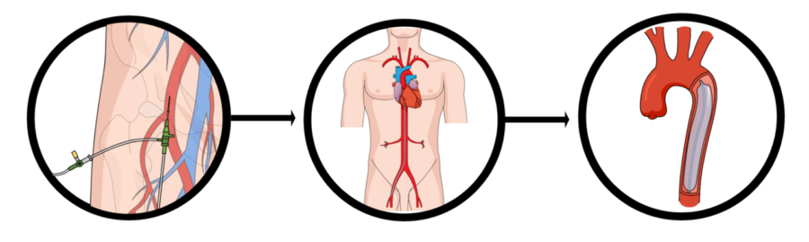
The IABP catheter which was prepared earlier is advanced over the guidewire and should be positioned with the tip of the balloon at the proximal aspect of the descending aorta, just distal to the left subclavian artery. Again, positioning should be confirmed with TEE or angiography.
The balloon is then flushed again with sterile saline and connected to a pressure transducer and to the IABP console which will include a gas line which provides the helium with which the balloon will be inflated. The IABP (and if applicable, the sheath) should be secured with sutures and then covered with sterile dressings.
Rule: The IABP should be secured at its proximal end (near the groin crease) and also at its distal end to ensure it cannot be accidentally advanced or withdrawn whilst the balloon is inflating.
Once the patient is stabilised, the balloon position should be checked on chest x-ray. The radio-opaque balloon tip should be visible on the CXR just below the aortic notch and just above the left hilum. Systemic heparinisation is not an absolute requirement when IABP is in situ but may be considered to limit possible thrombosis formation.
IABP Function
IABP systems have several potential triggers. They can be triggered by the patients ECG or by the observing pressure changes during each cardiac cycle (determined by the arterial line wave form).
When ECG triggered, the balloon inflates during the onset of diastole in the middle of the T wave and deflates just before the onset of systole at the peak of the R wave.
Poor ECG quality or arrhythmias can prevent IABP functioning when using ECG trigger.
When pressure triggered, the balloon will inflate following closure of the aortic valve determined by the dicrotic notch on the arterial line trace. It deflates just prior to aortic valve opening which is just before the upstroke on the trace. The program can assist every cardiac cycle or at a set timing. The commonest assistance patterns are one inflation per beat or one to two or one to three. The latter are commonly used when weaning a patient from IABP support when removal is being considered. Importantly, the balloon inflation can also be augmented in intervals of 10% with 100% representing maximal balloon inflation and 10% minimal. Less than 100% augmentation may be used for a patient who still requires the hemodynamic assistance of a balloon pump but is exhibiting signs of compromise due to the balloon, for example, limb ischemia.
Rule: Never leave an IABP in situ but not regularly inflating as this carries a high risk of thrombosis formation.
IABP Removal
When a patient is hemodynamically stable and no longer dependent upon IABP, it can be removed. To do this, have the patient lay supine with the ipsilateral leg slightly externally rotated. If a patient is on an inflated mattress, this should be inflated to maximal firmness. Again, much like prior to insertion, any factors associated with bleeding should be checked for normality. Following IABP insertion, especially if prolonged, patients may experience mechanical hemolysis resulting in anemia or thrombocytopenia and may require blood product transfusion immediately prior or during balloon removal.
Ensure you are wearing adequate PPE, that the patient’s bed is at a comfortable height for you to apply sustained pressure to the groin and that there is sterile gauze available. Dressings and securing sutures should be removed and the balloon should be stopped and set to standby on the console and the helium line disconnected. With one continuous movement, use gentle traction to remove the IABP (remove en-bloc with the sheath if a sheath has been used) in its entirety and provide immediate firm pressure to the area over the femoral artery puncture. Remember this may be a cm or two above the skin puncture site. Pressure should be maintained without interruption for at least 10 minutes. However, some advocate for longer. I personally hold 20 minutes of pressure prior to checking for hemostasis for all post-operative patients taking dual antiplatelet therapy or anticoagulation. The patient’s vitals should be monitored throughout and hemostasis checked prior to applying a pressure dressing. The patient’s blood work including hemoglobin and platelets should be checked a few hours later and the patient should maintain at least 2 hours bed-rest, supine, following IABP removal.
Complications of IABPs
The most worrisome complications of IABP use include acute limb ischemia (usually of the ipsilateral leg), severe bleeding, balloon leak and resultant helium embolus, thrombo-embolism formation and aortic dissection or rupture. Other complications which have been reported but are rare include visceral ischemia of gut organs. This would be due to IABP positioning too low and occlusion of the branches of the abdominal aorta by the balloon during inflation.
Patients who have smaller calibre peripheral arteries (particularly women) or those with aortic atheroma are particularly vulnerable to complications during insertion and removal. However, the SHOCK II trial found no significant differences in complications between patients who had IABP and those who did not.